|
A guide to spatialization of information to enhance perceptualization Jeff Sale |
||
 |
 |
 |
These are images of human bioelectric activity, such as electroencephalographic (EEG) and electrocardiographic (ECG) spatiotemporal isosurfaces rendered using the powerful AVS data rendering software on a Stardent Titan 3000 workstation (MIPS 3000 cpu). We did this work a few years ago and are belatedly getting it up on the web for you to see. Your comments and suggestions are welcomed.
What are these images? Click for an explanation.
|
|
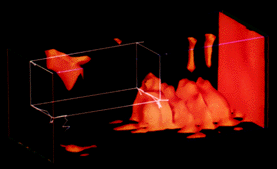
Alpha Bursts: The image to the left was created by calculating power in the alpha band (8-13 Hz) using a fast fourier transform (fft) on the eeg in Matlab, and outputing to AVS. |

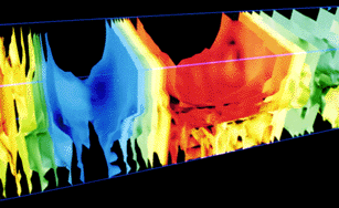
EEG Evoked
Potential: Created
from EEG data acquired using the P300 "oddball
paradigm". This image comprises roughly one second
of data. The blue region is N100, the red region is
P300.

EEG Evoked
Potential - Detail: Detail
of same data as above.
Alpha Bursts:
Created by
calculating power in the alpha band (8-13 Hz) using
a fast fourier transform (fft) on the eeg in
Matlab, and outputing to AVS.
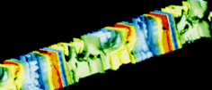
Raw Alpha
Waves:
These are alpha
waves acquired from a human subject, relaxed, eyes
closed, comprising roughly 3-4 seconds of activity.
This particular subject had especially clean and
strong alpha activity with eyes closed. This is apparent from the 'striped' red/blue pattern we see in the data. This is typical of very coherent (in phase) sinusoidal data.
Raw Alpha Waves -
Detail: EEG from
Electroconvulsive Therapy: Electrocardiogram
(ECG): EEG Correlation
Dimension:
This is a
detail of the same waves, focusing on a particular area comprising about one second of data. You can see that part of the eeg is slightly out of phase with the rest of the data.
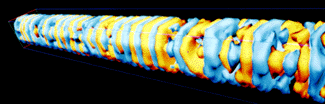
This data was
acquired intraoperatively from a patient undergoing
ECT. Note the "helical" pattern as time increases
from left to right.

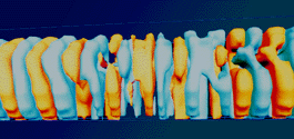
This is two
heartbeats worth of electrocardiographic data (ECG)
acquired by placing a 5x5 grid of electrodes on the
subject's chest. Note the repeating pattern of the
beats.
This is my
personal favorite. The others were easy to create
compared to this one. It took me a week to write
the code and the Stardent 2 weeks to calculate the
values. I wrote the algorithm to calculate the
correlation dimension, similar to the fractal
dimension, which is a measure of the complexity of
a system, or more generally, a measure of how a
system fills a space, real or abstract. The rest
was simple. The data consists of one minute of EEG,
the first 30 seconds the subjects eyes were open,
then they shut their eyes for the remaining 30
seconds. Time goes from left to right. As usual,
the visual cortex corresponds to the lower half of
the volume, which is where one would expect to see
the most significant change when the eyes are
closed, and this is in fact quite distinct.
It
is difficult to explain what these images are, but click
here to get a glimpse...